

A medical device program can lose months on imaging issues that were never in the original schedule. The sensor looked right on paper, but the optics failed under the actual working distance. The interface passed lab tests, then struggled in the final enclosure. That is why a medical imaging module integration guide matters early – not after the first prototype build.
For OEMs, system integrators, and engineering teams, imaging integration is not a single component decision. It is a system decision that affects image quality, thermal behavior, cable routing, latency, sterilization strategy, compliance planning, and production yield. In medical applications, small mistakes are expensive because performance is tied directly to clinical usability and product reliability.
What a medical imaging module integration guide should solve
A useful medical imaging module integration guide should help teams reduce technical rework before EVT and DVT. The real goal is not simply to select a camera module. It is to confirm that the module, optics, electronics, housing, and manufacturing process work together under the intended use conditions.
Medical products have very different imaging priorities depending on the use case. An endoscope module may prioritize compact diameter, illumination balance, moisture protection, and color consistency in close-range tissue imaging. A diagnostic reader may care more about fixed focus accuracy, distortion control, and repeatable exposure. A portable medical device may put power consumption and interface simplicity ahead of maximum resolution.
This is where many projects go off track. Teams often start with megapixel count and sensor brand, then discover that the harder integration work sits elsewhere. Optics, signal path stability, EMI control, and enclosure constraints usually decide whether the final image is clinically useful.
Start with the use case, not the sensor
The first integration decision is operational, not electrical. What does the camera need to see, at what distance, under what lighting, and for what type of user? A clinician using a handheld device has different expectations than a technician reviewing images on a workstation. The image pipeline should be designed around the decision being made from the image.
Resolution is often overemphasized. If the lens cannot resolve the sensor well at the needed field of view, higher resolution adds cost and data load without improving the image. Frame rate can also be misunderstood. For some medical imaging tasks, stable exposure and low noise matter more than high fps. For others, such as live scope guidance, latency and motion rendering are critical.
Depth of field, working distance, illumination geometry, and color response should be defined before module selection. If those requirements remain vague, integration becomes trial and error. That usually leads to multiple optical iterations, firmware changes, and enclosure modifications later.
Sensor and optics selection in medical imaging module integration
The sensor and lens need to be treated as a matched set. A strong sensor paired with the wrong optics will underperform in any environment, especially in medical use where close focus, narrow spaces, or reflective surfaces are common.
Sensor format affects more than image size. It influences lens options, module footprint, light sensitivity, and thermal layout. A larger sensor can improve image quality, but it also pushes enclosure size and optical stack depth. In compact medical devices, that trade-off is not always acceptable.
Lens choice should be driven by field of view, distortion tolerance, and target distance. In a device used for observation or inspection inside constrained anatomy or narrow channels, lens diameter and focus behavior may matter more than broad scene coverage. In external imaging devices, edge sharpness and color uniformity may take priority.
Auto focus is another area where it depends. In some systems, it improves usability. In others, it adds power draw, control complexity, and long-term reliability concerns. Fixed focus is often preferred when the device geometry and working distance are tightly controlled. That can simplify validation and make manufacturing more repeatable.
Electrical interface and data path decisions
MIPI, USB, DVP, and other interfaces each solve different problems. The right choice depends on processor architecture, required bandwidth, cable length, latency tolerance, and board design constraints.
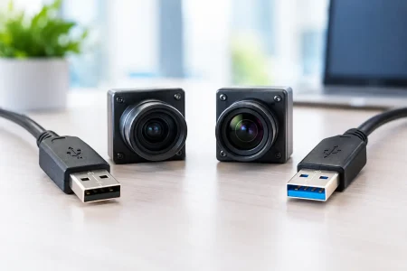
MIPI is common in embedded medical devices where compact board layout and direct processor integration are important. It supports high data throughput but requires tighter signal integrity control and closer attention to PCB stack-up, cable design, and connector quality. USB modules are easier for rapid prototyping and system compatibility, especially for host-based medical systems, but they can introduce power and cable considerations that become more visible in finished products.
DVP can still fit certain lower-complexity designs, but teams should confirm long-term processor compatibility and image bandwidth headroom. A design that works at prototype stage may become constrained when image processing features are added later.
The data path should be reviewed end to end. Sensor output, bridge processing, ISP behavior, host compatibility, compression method if any, and display latency all shape user experience. A good image at the sensor is not enough if noise enters through the cable, if the ISP handles color poorly, or if dropped frames appear under sustained operation.
Mechanical integration is where many delays begin
Camera module integration often fails at the mechanical level before the electronics are fully debugged. Stack height, connector position, flex routing, adhesive choice, tolerance build-up, and shock behavior all affect the final result.
Medical devices frequently have tighter packaging constraints than industrial equipment. The camera may sit near LEDs, batteries, motors, or shielding structures. Every nearby component can affect heat, noise, and optical performance. Even slight module tilt can shift focus across the image or create calibration inconsistency between units.
If the product requires sterilizable or disposable elements, the mechanical strategy becomes even more important. Material selection, sealing approach, and assembly process must support both image quality and product lifecycle requirements. There is no universal answer here. A reusable device and a single-use device will push integration decisions in very different directions.
Power, heat, and image stability
Medical imaging systems are expected to perform consistently over time, not just during a short bench test. That makes power integrity and thermal management core integration issues.
Unstable power can show up as image noise, intermittent detection failures, or startup inconsistency. Heat can shift sensor behavior, alter color response, and raise noise levels during long operating sessions. Small devices are especially vulnerable because there is less room for thermal spreading and less tolerance for hot spots.
Engineering teams should test the module in realistic duty cycles. Continuous streaming, LED illumination load, enclosure closure, and battery-powered operation can reveal conditions that do not appear in open-air development setups. It is much better to identify these limits before tooling is locked.
Validation should reflect clinical use, not only lab conditions
A strong validation plan looks beyond resolution charts. Those are useful, but they are not enough. Medical imaging modules should be evaluated under real lighting conditions, real target materials, realistic working distances, and likely contamination or moisture exposure if the product environment demands it.
Image quality should be judged against the application. White balance, noise, flare control, shading, color repeatability, and low-light behavior may each matter differently depending on the device. If software will process the image for recognition, measurement, or enhancement, validation should include that pipeline too.
Manufacturing teams should also care about repeatability. Can the optical stack be assembled with stable focus across production lots? Can incoming inspection catch variation early? Can calibration be standardized without slowing line throughput? These are commercial questions, but they are also engineering questions.
Supplier capability is part of the integration plan
For medical products, the module supplier is not just a parts vendor. The supplier affects sample speed, design revisions, production consistency, and troubleshooting efficiency. If customization is required, the supplier’s engineering depth matters as much as the initial spec sheet.
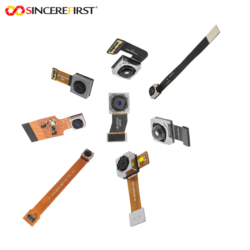
Teams should ask practical questions. Can the supplier adapt lens, connector, board shape, flex length, and housing constraints? Can they support quick sample turns for iterative testing? Can they scale from prototype quantities to mass production with controlled quality? Those answers influence schedule risk directly.
This is one reason many OEMs prefer a manufacturing partner with both standard module experience and custom imaging development capability. A supplier that understands optical tuning, interface behavior, and production process control can help prevent redesign loops. For companies building commercial medical devices, that support often saves more time than any single hardware feature.
A practical medical imaging module integration guide for faster execution
If you want integration to move faster, define the image task first, lock the mechanical envelope early, and review the full signal path before selecting the module. Then validate under sustained operating conditions, not just ideal bench tests. The teams that do this well are not guessing less because the product is simpler. They are guessing less because the integration plan is tighter.
SincereFirst works with OEM and device teams that need that level of control – from compact endoscope modules to customized medical imaging configurations built for manufacturability and stable supply. The most efficient programs usually start with clear constraints, quick engineering feedback, and a supplier that can support both prototype refinement and volume production.
If your current design review is still centered on megapixels alone, pause there. The better question is whether the full imaging module will perform inside the real product, under the real workflow, at the volume you plan to ship.