
A microscope can have excellent optics and still produce poor inspection results if the camera is specified incorrectly. The best cameras for microscopes are not defined by resolution alone. They must match the objective lens, field of view, illumination, mounting constraints, data interface, and the decision the end user needs to make from each image.
For OEM device teams, this distinction matters early. A camera that looks impressive in a desktop demonstration may create color inconsistency, motion artifacts, thermal issues, or bandwidth limits after it is integrated into a clinical instrument, industrial inspection station, or compact automated analyzer. The right camera is the one that produces repeatable, usable image data at the required scale.
What Makes a Microscope Camera the Right Choice?
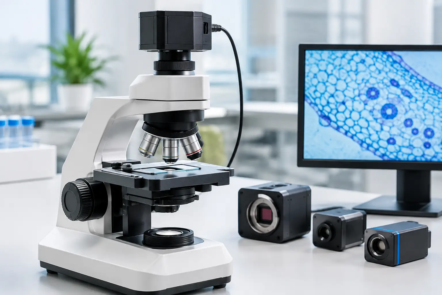
Microscope imaging is an optical system problem, not simply a sensor selection problem. The objective determines magnification and numerical aperture. The tube lens, relay optics, and camera adapter determine how the image circle reaches the sensor. The camera then converts the available optical information into digital data.
That chain creates a practical limit: adding more megapixels does not recover detail that the objective, optical path, or illumination cannot resolve. Conversely, an undersized sensor or large pixel pitch can waste useful optical resolution. Engineering teams should begin with the sample size, required smallest feature, working distance, and desired field of view before requesting a camera module.
A camera also needs to fit the operating workflow. A pathology viewer may prioritize accurate color and high-resolution still capture. A live cell imaging platform may need high sensitivity and low phototoxicity. A production inspection system may require a global shutter, deterministic triggering, and rapid USB 3.0 or MIPI data transfer. These are fundamentally different requirements, even when all three systems use microscopes.
Best Cameras for Microscopes by Application
Brightfield and general laboratory imaging
For brightfield documentation, education, and routine laboratory imaging, color sensors with 2 MP to 12 MP resolution are common starting points. A sensor with good color reproduction, low fixed-pattern noise, and stable automatic or manually controlled white balance can be more valuable than an unnecessarily high resolution option.
Pixel size should be evaluated alongside the microscope’s effective magnification. Smaller pixels can improve sampling when the optics support it, but they also collect less light. Under modest LED illumination, that can increase gain, noise, or exposure time. For a general-purpose instrument, a balanced sensor with reliable color response often delivers better day-to-day results than a specification-heavy sensor pushed beyond its light budget.
Fluorescence and low-light imaging
Fluorescence microscopy changes the priorities. Signal levels are often limited, while preserving sample integrity may require low illumination intensity and short exposure. Sensitivity, quantum efficiency, read noise, dark current, and cooling strategy become central selection criteria.
Monochrome cameras are frequently preferred when maximum sensitivity is required because they do not use a color filter array. They capture more usable signal per pixel and can be paired with controlled excitation and emission filters. Color cameras remain appropriate for certain multichannel or documentation tasks, but their performance must be judged in the actual optical configuration rather than under standard room-light test conditions.
For compact instruments, sensor heat and module heat deserve careful attention. A camera module placed close to a sensitive optical assembly can affect noise performance, mechanical stability, and long-duration reliability. Housing design, heat transfer, and cable routing should be reviewed as part of the imaging design.
Live microscopy and moving samples
High frame rate is critical when imaging moving organisms, fluid behavior, conveyor-fed samples, or motorized scanning stages. In these applications, a global shutter camera is usually the safer choice. It exposes every pixel at the same moment, which avoids geometric distortion caused by motion during readout.
Rolling shutter sensors can still be effective for stationary samples and cost-sensitive instruments. They offer broad sensor availability and can provide strong image quality. However, they need validation with the intended stage motion, vibration level, strobe illumination, and exposure time. Motion blur and rolling shutter skew are system-level issues, not defects that can always be corrected later in software.
Industrial and automated inspection microscopy
Automated visual inspection demands consistency more than visually pleasing images. The camera should support fixed exposure, gain, white balance, trigger behavior, and image output settings so an inspection algorithm receives stable data from unit to unit.
Monochrome cameras are often selected for measurement, edge detection, and defect classification because they eliminate color interpolation and simplify illumination control. Color imaging is justified when material hue, contamination, coating variation, or label identification is part of the inspection target. In either case, hardware triggering and controllable I/O can be decisive when the microscope is part of a larger automation cell.
Sensor Specifications That Actually Matter
A useful camera specification begins with five connected questions:
- What smallest feature must be detected or measured?
- How much field of view is required in one frame?
- How much light is available at the sensor plane?
- Is the subject stationary during exposure?
- How quickly must images be transferred, processed, and stored?
Resolution answers only part of the first two questions. A 5 MP sensor can be a better fit than a 20 MP sensor if its active area matches the microscope image circle and its pixel size supports the optical resolution. An oversized sensor may create vignetting or force an adapter design that adds cost and alignment risk.
Dynamic range is equally important for specimens with bright highlights and dark regions. A higher dynamic range sensor helps preserve information across the image, reducing clipped bright areas or crushed shadow detail. Bit depth should be considered with the full acquisition chain. Capturing 10-bit or 12-bit output is valuable only when the interface, processor, storage, and image-processing software can retain and use that data.
Noise performance is not one number. Read noise matters in low-signal conditions, while dark current becomes more relevant during long exposures or elevated temperatures. Fixed-pattern noise can affect quantitative measurements if it is not controlled through sensor selection, calibration, and image correction.
Choose the Interface Before Finalizing the Camera
The interface determines more than connector type. It affects bandwidth, cable length, host processor selection, software architecture, power design, and manufacturability.
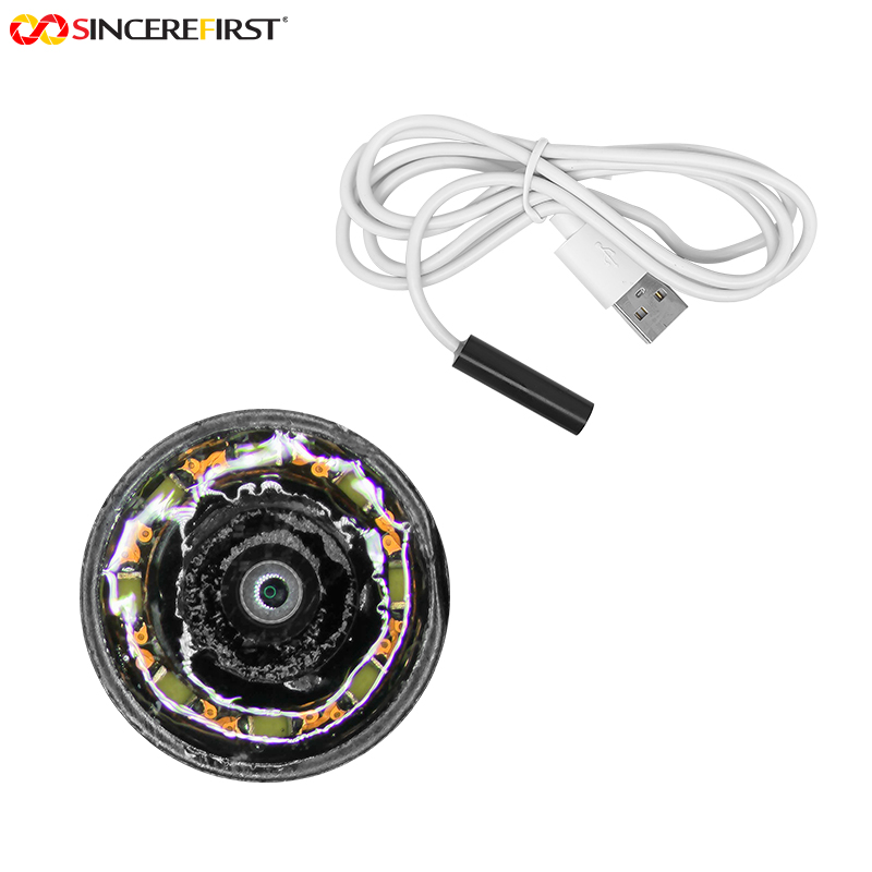
USB 2.0 camera modules remain practical for moderate-resolution imaging and straightforward plug-and-play integration. USB 3.0 supports higher frame rates and larger image streams, making it a strong option for laboratory instruments and industrial workstations with a nearby host computer. UVC compatibility can shorten software integration for applications that benefit from standard camera support, although it may offer less control than a dedicated SDK or custom firmware approach.
MIPI CSI-2 is well suited to compact embedded products using application processors or system-on-chip platforms. It reduces module size and can offer efficient high-speed image transfer, but signal integrity, flex cable length, board layout, and operating system driver support must be planned carefully. DVP interfaces can still suit lower-speed embedded designs, particularly where simplicity and legacy processor compatibility matter.
For production programs, avoid specifying the interface in isolation. Confirm the complete path from sensor output to display, storage, AI inference, or inspection software. Frame drops that appear only under full system load are expensive to discover after mechanical tooling is complete.
Mechanical Integration and Optical Matching
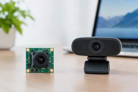
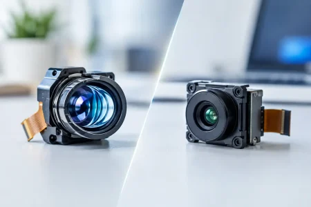
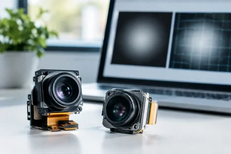
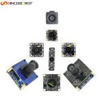
Many microscope cameras are attached through C-mount adapters, but an OEM system may need a smaller board camera, custom FPC camera module, or integrated optical assembly. The decision depends on available space, service requirements, target cost, and whether the product will use standard microscope hardware or a purpose-built optical stack.
Sensor size must match the projected image circle. If the sensor is too large, image corners may darken. If it is too small, the system crops the field of view. Adapter magnification also changes the effective pixel size at the specimen plane, which directly affects sampling and measurement accuracy.
Mechanical tolerances deserve the same attention as sensor specifications. Focus drift, tilt, vibration, connector retention, and thermal expansion can reduce image quality even when the optics and camera are individually well designed. For medical, industrial, and field-deployed equipment, validate those factors across temperature, shock, handling, and extended operating cycles.
Build for Calibration, Supply Stability, and Scale
A commercial microscope camera program should include calibration planning from the beginning. Depending on the application, this can include white balance calibration, shading correction, defective-pixel correction, geometric calibration, and color target verification. Inspection and measurement systems may also require traceable calibration procedures and controlled software versions.
Supply continuity is another engineering requirement. Sensor availability, lens sourcing, connector life, firmware management, and test procedures should be assessed before volume release. A camera module that meets performance targets but changes unexpectedly during a product lifecycle creates qualification work and customer risk.
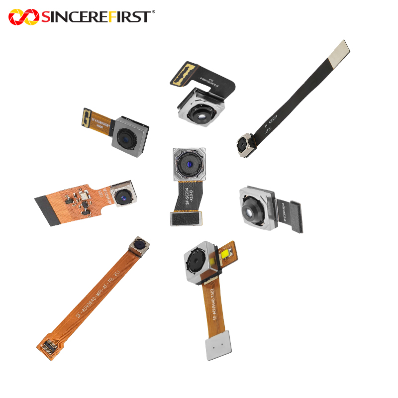
For teams developing specialized microscope instruments, a manufacturer with module, optical, firmware, and production engineering capability can reduce handoff delays. SincereFirst supports this approach with standard and customized camera modules for embedded imaging programs where mechanical dimensions, sensor selection, interfaces, and image tuning must be controlled together.
The most effective next step is to test candidate cameras in the real optical path, under the real illumination, with representative samples and production-intent software. That evidence will identify the camera that supports a dependable instrument, not merely the one with the most attractive datasheet.