
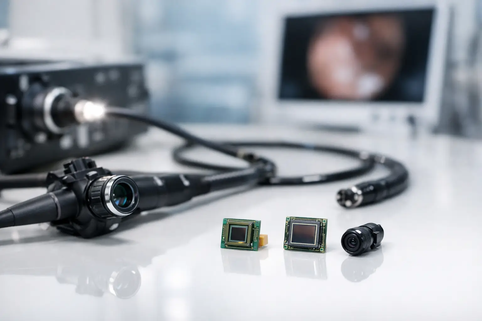
A sensor decision made at the camera-module stage can determine whether a medical endoscope delivers clinically useful detail or disappointing images in the darkest, tightest anatomy. The question is not simply which sensor has the highest resolution. Which sensor fits medical endoscopes depends on the distal tip diameter, optical design, illumination level, frame-rate requirement, image-processing pipeline, sterilization strategy, and target procedure.
For most new medical endoscope programs, a CMOS image sensor is the practical starting point. CMOS technology supports compact packaging, low power consumption, high frame rates, and efficient integration with MIPI or other digital interfaces. However, the best sensor for a 0.9 mm disposable scope is not the best choice for a larger reusable laparoscope or a specialty device that requires fluorescence imaging. Engineering teams should select the sensor as part of a complete imaging system, not as an isolated specification.
Which Sensor Fits Medical Endoscopes by Scope Size?
The outer diameter of the insertion tube and distal tip creates the first hard limit. In ultra-slim endoscopes, the image sensor, lens, LEDs, wiring, and mechanical enclosure compete for fractions of a millimeter. A sensor with an attractive data sheet may be physically impossible to package once lens height, bonding area, heat management, and assembly tolerances are included.
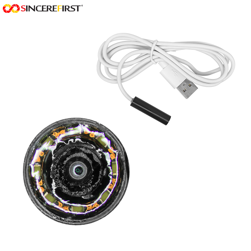
For endoscope camera modules below approximately 2 mm in diameter, sensor size is usually the dominant constraint. Small-format CMOS sensors can support applications such as ENT inspection, dental imaging, disposable diagnostic devices, and narrow-channel medical instruments. The trade-off is reduced light capture. Very small pixels and a small optical format demand carefully matched optics and sufficient illumination to maintain acceptable signal-to-noise performance.
As the module diameter increases into the 3 mm to 10 mm range, designers gain more freedom to select larger optical formats, higher resolutions, and lenses with better low-light performance. This is where 720p, 1080p, or higher-resolution CMOS sensors become more feasible. A larger sensor can improve image quality, but it also requires an optical system that covers its active area and preserves sharpness across the field of view.
Do not select by sensor package dimensions alone
The active array, package width, package height, lens mount approach, flex routing, and distal housing all matter. A module that fits in a CAD layout can still become difficult to manufacture at volume if assembly margins are too tight. For medical OEMs, repeatable alignment and yield are as important as achieving a one-off prototype.
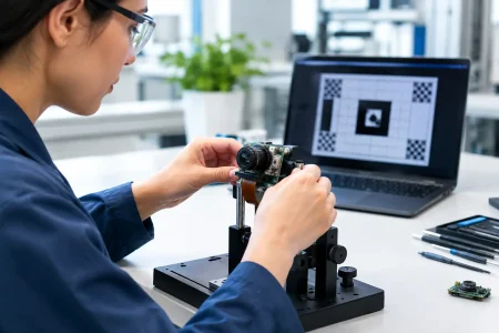
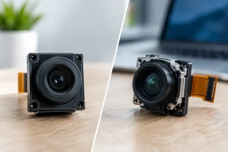
A capable endoscope module supplier should validate the full stack: sensor package, lens geometry, LED placement, FPC design, sealing method, and finished distal diameter. This prevents late-stage redesign when a nominally suitable sensor cannot meet the mechanical target in production.
Start With Clinical Image Requirements, Not Resolution
Higher resolution is valuable only when the optics, illumination, display, and processing chain can preserve that detail. A 2 MP sensor may provide better clinical usability than a poorly illuminated 5 MP sensor with excessive noise, motion blur, or lens-edge softness.
For general visualization, 720p or 1080p can provide an effective balance of image detail, bandwidth, power, and cost. Higher resolution may be justified when the procedure requires digital zoom, close inspection of fine tissue structures, image recording, or large-screen viewing. It can also introduce higher processing loads, greater storage requirements, and more demanding thermal management.
Pixel size deserves equal attention. Larger pixels generally capture more photons, which helps in low-light conditions and can reduce noise. This matters in endoscopy because illumination is limited by available distal-tip space, LED heat, power delivery, and patient safety requirements. Small pixels can support high resolution in a compact sensor, but they often need stronger lighting and more advanced image processing to produce clean images.
Color performance is another practical differentiator. Medical users need consistent color rendering to interpret tissue appearance. Sensor color filter characteristics, white-balance control, LED spectral output, lens transmission, and image signal processor tuning all influence the final image. A sensor cannot be evaluated properly without representative optics and illumination.
Shutter Type Matters When Motion Is Fast
Most compact CMOS sensors use a rolling shutter, which reads the image line by line. Rolling shutter sensors are common in endoscope modules because they offer strong resolution, sensitivity, availability, and cost performance in small formats. They are usually suitable for many inspection and visualization procedures when illumination and frame rate are well controlled.
A global shutter captures all pixels at the same moment. It can reduce distortion caused by rapid movement of the scope, tissue, instrument, or patient. This is particularly relevant when motion fidelity is critical or when the endoscope works alongside moving tools. The trade-off is that global shutter sensors may offer fewer choices in very small packages, different sensitivity characteristics, or a higher system cost.
The correct choice depends on the expected motion profile. If a device will be used mainly for controlled inspection, rolling shutter may be the more efficient design choice. If rapid movement or motion measurement is central to the procedure, evaluate global shutter options early rather than attempting to correct distortion later with software.
Match the Sensor Interface to the Host Architecture
A medical endoscope camera module must communicate reliably with the control board, processor, or external display system. The sensor interface affects cable design, latency, electromagnetic compatibility, power consumption, and integration effort.
MIPI CSI-2 is a common choice for compact digital camera modules connected to modern embedded processors. It supports high data rates and is well suited to short, controlled interconnects. DVP interfaces can simplify integration with certain legacy or cost-sensitive platforms but may have bandwidth limitations at higher resolutions. USB camera modules are useful when the medical device architecture benefits from plug-and-play UVC connectivity, though the module size and cable arrangement may not suit every distal-tip design.
For a long insertion tube, the raw sensor interface may not be practical over the full cable distance. The system may require a serializer-deserializer architecture, a dedicated processing board, or image transmission design that accounts for signal integrity. Sensor selection should therefore follow a bandwidth calculation that includes resolution, frame rate, bit depth, lane count, and overhead – not a simple comparison of megapixels.
Low Light, LEDs, and Heat Form One Design Problem
The sensor, illumination system, and thermal design are tightly connected. Increasing LED output can improve exposure, but it also creates heat at the distal end. Increasing sensor gain can brighten an image, but excessive gain increases noise and reduces fine detail. Slowing the exposure improves photon collection, but it can cause motion blur.
The practical objective is a balanced exposure system: a sensor with suitable sensitivity, an optical design with efficient light transmission, LEDs selected for color quality and output, and image processing tuned to the intended procedure. This balance often matters more than selecting the largest available sensor.
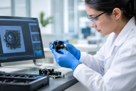
A prototype should be tested in representative optical conditions rather than on a bright laboratory target. Evaluate color, noise, highlight control, shadow detail, autofocus or fixed-focus behavior, and image stability under expected working distances. For a fixed-focus endoscope, depth of field is especially important. A sensor-lens combination that looks sharp at one distance may fail to provide useful clarity across the clinical working range.
Medical Product Development Requires Supply Stability
For commercial medical devices, a sensor must be more than technically capable. It must be available through the product lifecycle, supported by controlled specifications, and compatible with a repeatable manufacturing process. A consumer-oriented sensor with uncertain lifecycle status can create costly redesign risk after a device has entered verification or regulatory submission.
Engineering and procurement teams should confirm sensor lifecycle expectations, lot consistency, interface documentation, image tuning support, and second-source or redesign planning where appropriate. They should also define incoming inspection criteria for dead pixels, image uniformity, color consistency, and cosmetic requirements. These details protect product quality as production scales.
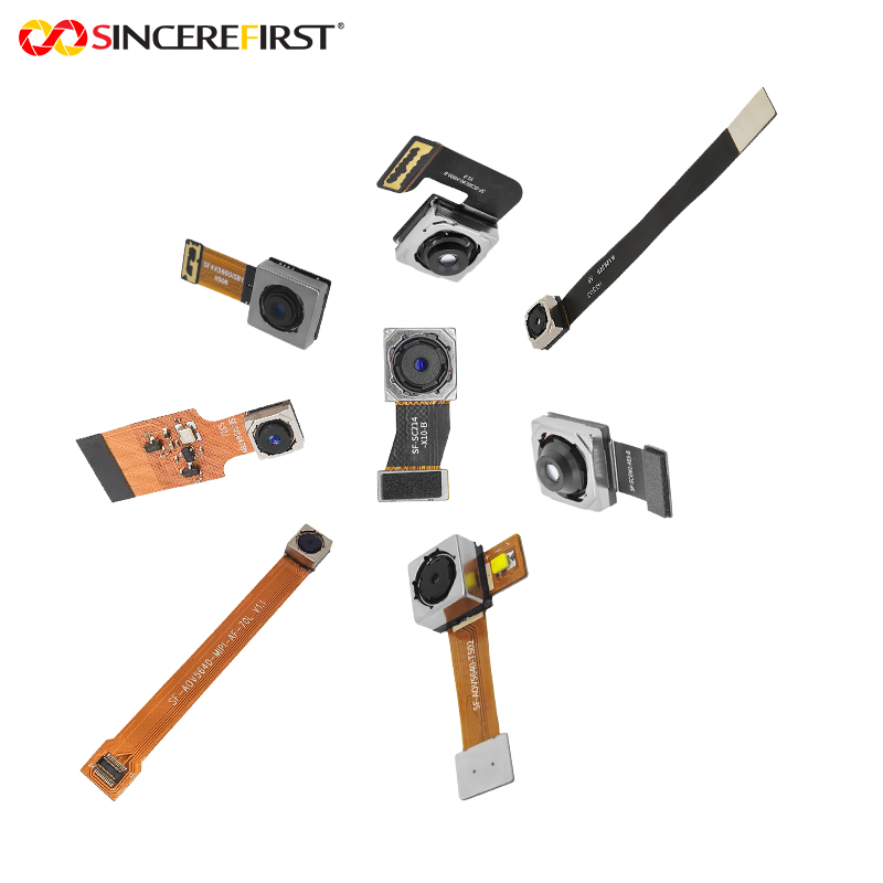
SincereFull, the endoscope camera module brand of SincereFirst, supports ready-made and customized modules from 0.9 mm to 10.0 mm in diameter. The most effective projects begin with a shared definition of the procedure, mechanical envelope, image target, interface, and anticipated production volume. That allows sensor, optics, illumination, and manufacturing decisions to be resolved together rather than in separate stages.
A Practical Sensor Selection Path
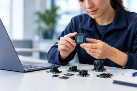
Begin by defining the non-negotiable requirements: distal diameter, required field of view, working distance, target resolution, frame rate, illumination limits, and host interface. Next, compare candidate CMOS sensors based on optical format, pixel size, sensitivity, shutter type, power, and package dimensions. Then build and test a complete module with the intended lens, LEDs, cable, and processor.
The final decision should come from images captured under realistic use conditions and from a manufacturing review that confirms assembly feasibility, quality controls, and supply continuity. The right sensor is the one that produces dependable clinical images inside the required form factor while remaining manufacturable at the quality and volume your medical device program requires.