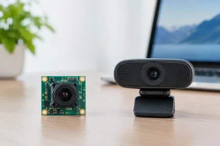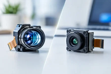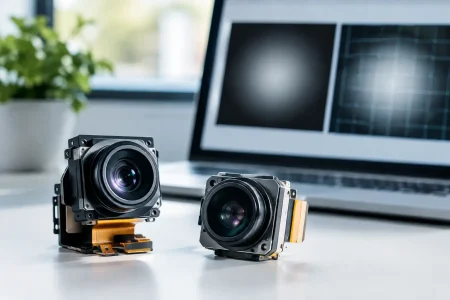

A medical endoscope can be only a few millimeters in diameter, yet its imaging chain must deliver clinically useful detail under conditions that are unforgiving: confined anatomy, reflective tissue, variable working distance, heat limits, repeated cleaning, and strict device-validation requirements. For OEM teams, medical endoscopy is not simply a matter of selecting the smallest available camera module. It is a system-level engineering decision that affects image quality, usability, manufacturability, regulatory documentation, and long-term supply.
The camera module is central to that decision, but it cannot be evaluated in isolation. Sensor performance, lens design, illumination geometry, cable construction, image signal processing, and mechanical packaging all influence what a clinician sees. A module that looks strong on a bench chart can still underperform once it is integrated into a finished scope, catheter, or handheld diagnostic device.
Medical Endoscopy Starts With the Clinical Use Case
The first specification should be the procedure, not the resolution. A rigid arthroscope, a disposable ureteroscope, a single-use ENT device, and a capsule-style inspection platform can all have very different imaging priorities. Some require a wide field of view to maintain orientation. Others need close-focus performance for tissue inspection. Some need a forward-viewing camera, while others require a side-view or angled-view configuration.
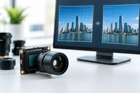
Working distance is especially important. A lens optimized for sharp imaging at 10 mm may not provide the required detail at 30 mm. Depth of field, distortion, and illumination falloff must be considered together. In a narrow cavity, a very wide-angle lens can improve situational awareness but may introduce edge distortion that complicates clinical interpretation or software measurement.
Resolution also needs context. More pixels can support detail, digital zoom, and larger displays, but higher resolution can increase data bandwidth, processing demand, power consumption, and thermal load. For a compact disposable device, a balanced sensor and optical design may deliver more practical value than a high-resolution sensor constrained by poor lighting or limited transmission bandwidth.
Selecting the Endoscope Camera Module
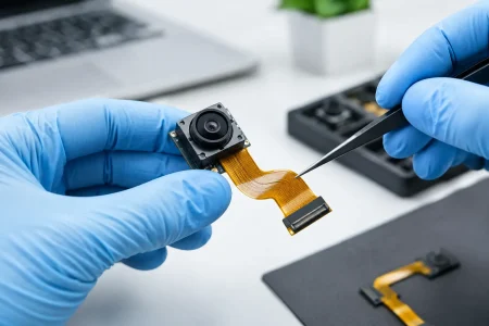
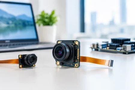
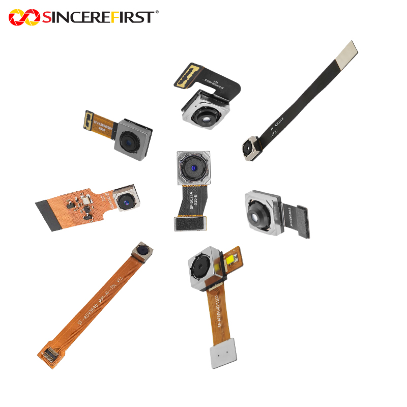
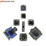
For OEM and ODM programs, module selection normally begins with size, interface, image requirements, and integration constraints. Diameter is often the first limiting factor. Ready-made endoscope camera modules are available across a wide range, including ultra-compact designs from approximately 0.9 mm through larger 10.0 mm configurations. However, diameter alone does not determine suitability. The usable optical aperture, cable routing, lens stack, and housing tolerance must all fit the finished device architecture.
Sensor size, sensitivity, and pixel performance
A small sensor is often necessary for a narrow insertion tube, but the trade-off is reduced light collection. Pixel size, quantum efficiency, read noise, dynamic range, and low-light performance become more consequential as module dimensions shrink. Medical scenes frequently contain both bright specular highlights and darker recessed areas, making dynamic range and exposure control essential.
A sensor with strong low-light behavior can reduce the illumination burden, which may help manage heat and power. Yet the sensor should still be evaluated with the selected lens and LEDs, not only from a datasheet. Image quality depends on the entire optical path, including lens transmission, color filter response, protective windows, and the optical effect of any distal cover.
Interface choice affects the complete device
MIPI CSI-2 is often selected for compact embedded platforms because it supports high-speed image transfer with a small connector footprint. USB camera modules can simplify development for PC-based systems, external displays, and certain portable diagnostic products. DVP remains relevant in legacy or cost-sensitive architectures, while custom board-to-board and FPC designs are common where internal space is tightly controlled.
The right interface depends on the host processor, cable length, target frame rate, power budget, and software stack. A technically capable module can create unnecessary risk if its driver support or signal routing requirements do not align with the host platform. Early coordination among optical, electrical, firmware, and mechanical teams reduces late-stage redesign.
Integrated versus separate camera architectures
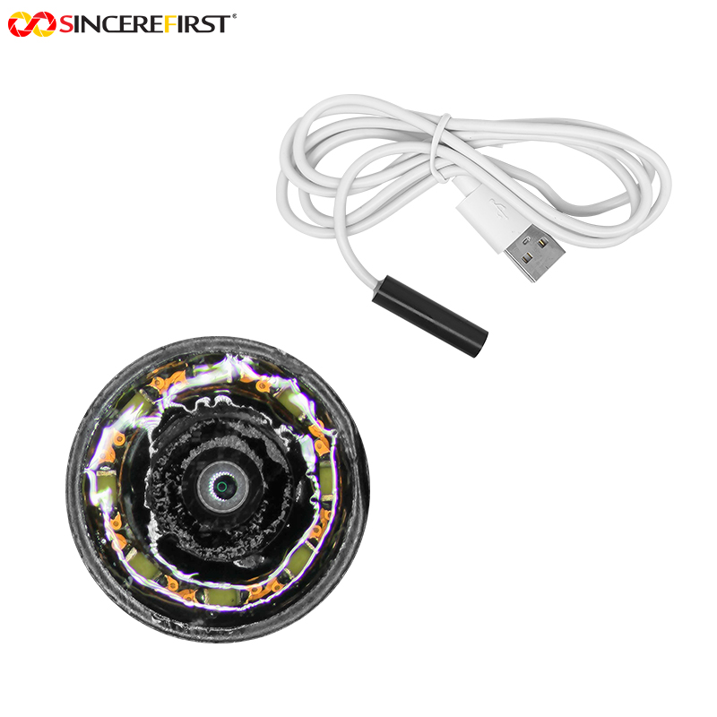
An integrated endoscope camera module combines the sensor, lens, and related electronics into a compact assembly. This approach can reduce integration effort and improve consistency when a standard configuration fits the application. A separate architecture can offer more flexibility in locating electronics away from the distal tip, which may help manage diameter, thermal performance, or serviceability.
Neither approach is automatically superior. An integrated design is often advantageous where development speed and compact packaging are primary requirements. A separate design may be better when the device requires a specific distal profile, longer cable routing, or a custom handle and processing architecture.
Illumination Is Part of Image Quality
Poor illumination is one of the fastest ways to compromise an otherwise capable imaging system. In medical endoscopy, light must reach the target evenly without excessive hot spots, shadows, glare, or heat at the distal end. LED selection, placement, optical diffusion, drive current, duty cycle, and thermal conduction all matter.
Color quality matters as well. Inadequate spectral output can make tissue tones appear unnatural, while overly aggressive image processing can mask the underlying problem rather than correct it. OEM teams should assess color rendering under the intended illumination source and display pipeline. White balance, auto exposure, gamma, denoising, and sharpening should be tuned to preserve useful clinical detail, not merely create a visually dramatic image.
For small-diameter devices, the illumination trade-off is sharper. There is limited physical space for LEDs, heat dissipation, and optical separation between light sources and the camera. This is where custom mechanical design and optical validation provide a meaningful advantage over selecting components independently.
Reliability Must Be Designed Into the Module
Medical-device buyers need repeatable performance across samples, lots, and production years. A camera module that meets image targets in a prototype but varies significantly in focus, color response, or connector reliability is not production-ready.
Critical reliability considerations include cable flex life, connector retention, solder-joint strength, lens alignment stability, moisture resistance, and tolerance to the intended cleaning or sterilization process. Reusable devices may face repeated exposure to chemicals, temperature cycles, or sterilization environments. Single-use devices have different priorities, often centered on cost control, assembly yield, shelf-life stability, and consistent performance at volume.
The distal assembly deserves particular attention. A minor shift in lens position can affect focus. A weak adhesive selection can create failure under temperature or humidity stress. A protective window can introduce reflections, contamination traps, or optical distortion if it is not designed as part of the lens system. These are manufacturing details, but they become clinical-image and product-reliability issues in the field.
Verify Image Performance in the Finished Device
Supplier qualification should go beyond reviewing sample images. OEM teams should establish objective acceptance criteria before pilot production. These may include resolution at defined working distances, field of view, depth of field, distortion, color consistency, illumination uniformity, frame rate, latency, dead-pixel limits, and cosmetic requirements.
The test environment should reflect actual use as closely as possible. Evaluate images through the final distal cover, cable, illumination system, and host processor. Assess the device on the intended display and with the final image-processing settings. If a scope will be used in a fluid-filled environment or behind a protective sheath, those conditions should be represented in validation testing.
Manufacturing controls are equally important. Ask how focus is set and verified, how lens alignment is controlled, what incoming inspection is applied to sensors and optics, and how traceability is maintained. For scaled programs, the supplier’s ability to support engineering samples, design changes, pilot builds, and stable volume production can be as important as initial module performance.
Customization Should Solve a Defined Constraint
Custom medical endoscopy development is most effective when it is tied to a clear technical need: a required diameter, a nonstandard viewing angle, a specific cable length, a host interface, a waterproofing target, or a particular illumination arrangement. Customization without defined acceptance criteria can extend development without improving the final device.
SincereFull, the endoscope module brand of SincereFirst, supports both ready-made and customized endoscope camera module configurations for medical and industrial applications. For device teams, the practical value of a capable manufacturing partner is not only access to component options. It is the ability to convert clinical and mechanical requirements into a controlled, producible imaging assembly with fast samples and a path to volume supply.
The strongest medical endoscopy programs treat imaging as a coordinated design discipline from the first prototype onward. Specify the clinical task, validate the full optical chain, and select a module partner that can maintain the same precision when the project moves from engineering bench to production line.