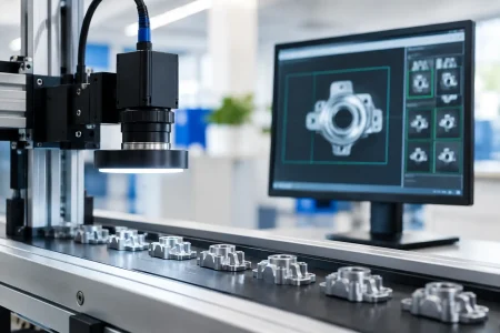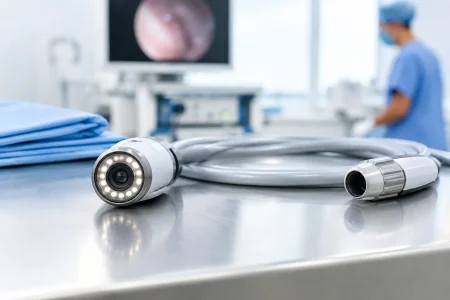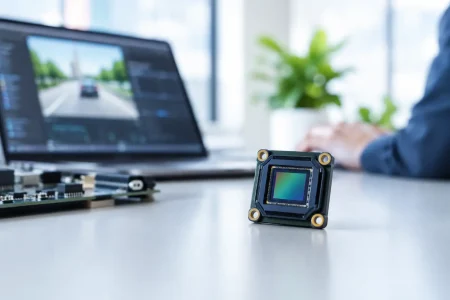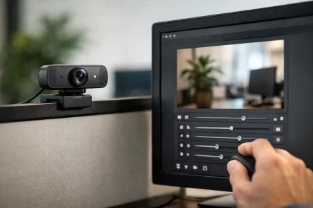

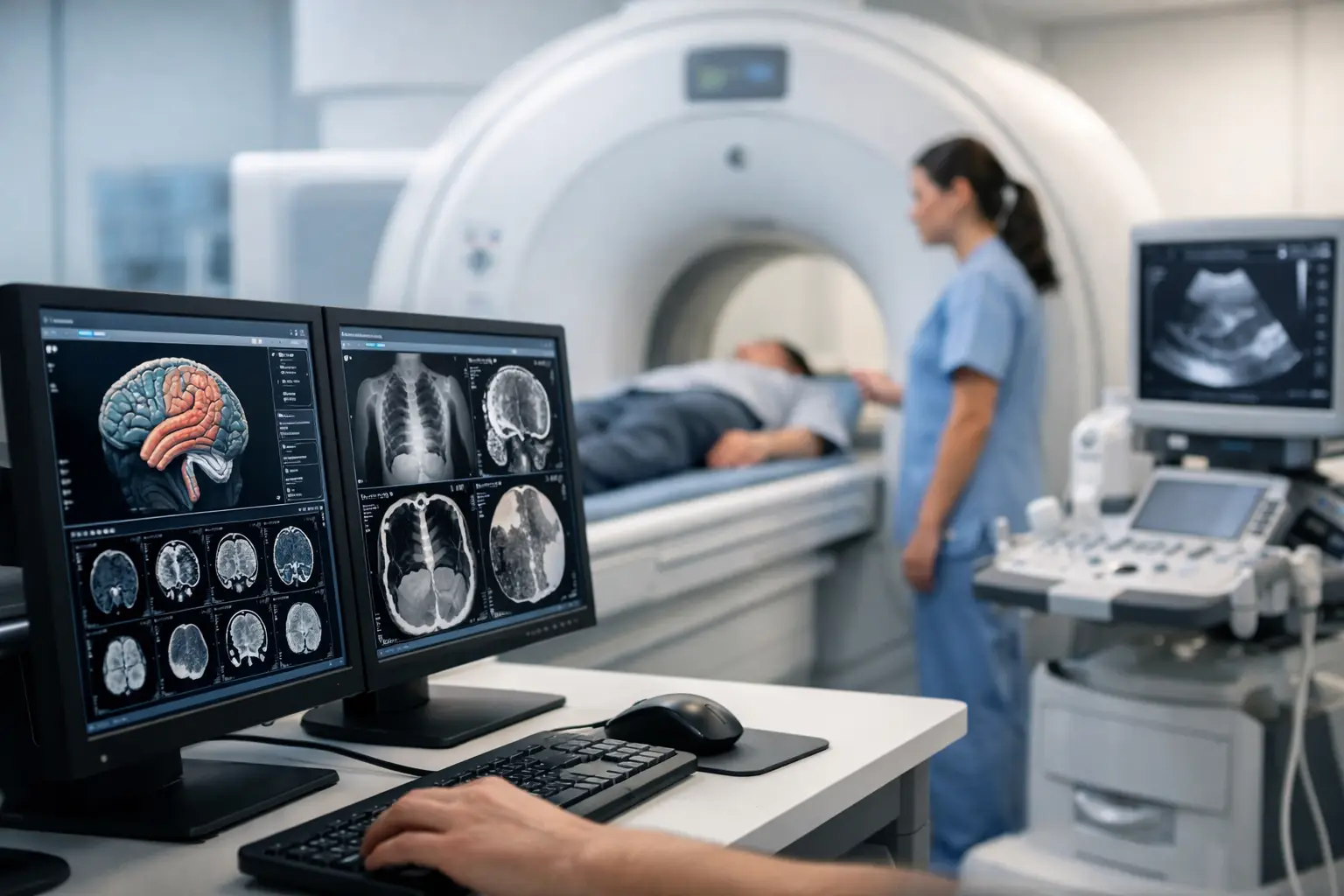
A medical imaging module is not selected by resolution alone. In an endoscope, handheld diagnostic device, dental camera, or point-of-care instrument, the top medical imaging module features determine whether the final product delivers usable clinical detail, fits its mechanical envelope, maintains stable output, and can be manufactured consistently at volume.
For OEMs, the right specification begins with the clinical task. A module intended to document skin conditions has different requirements from one designed to image small anatomical structures in a narrow, illuminated cavity. The sensor, optics, lighting, interface, housing, and production controls must operate as one engineered system.
Top medical imaging module features to evaluate
Image quality at the working distance
Resolution matters only when it produces meaningful detail at the device’s actual working distance. A higher-resolution sensor can support cropping, digital zoom, and fine-detail inspection, but it also increases data throughput, processing demand, power consumption, and storage requirements. For many compact medical devices, a well-matched 1080p or 2MP module can deliver more useful results than a high-pixel-count sensor paired with unsuitable optics.
Ask for image samples captured at the expected distance, field of view, illumination level, and target surface. Center sharpness alone is not enough. Evaluate edge performance, distortion, color consistency, noise, motion artifacts, and the ability to maintain focus across the relevant depth range. These factors directly affect whether clinicians can interpret the image without repeated repositioning.
Sensor size and pixel size also require a practical trade-off. Larger pixels generally collect more light and can improve low-light performance, while a smaller sensor may be necessary for a slim probe, disposable attachment, or tightly packed handheld enclosure. The preferred option depends on the optical path and intended procedure, not on a single headline specification.
Optical design and depth of field
The lens is a primary determinant of imaging performance. Field of view must cover the clinical area without introducing distortion that interferes with interpretation. In close-range imaging, depth of field is often equally important. A module that looks sharp at one fixed distance may become unusable when normal hand movement shifts the target by only a few millimeters.
Fixed-focus optics are compact, cost-effective, and stable when the working distance is controlled. Autofocus can improve flexibility for external examination devices or general-purpose diagnostic cameras, but it adds mechanical complexity, power demand, startup time, and potential failure modes. For many endoscopic and miniature medical applications, precise fixed-focus calibration is the more reliable engineering choice.
Lens selection should also account for glare, reflections, and surface texture. Anti-reflective coatings, appropriate aperture settings, and careful illumination geometry can improve contrast on wet, glossy, or uneven tissue surfaces. These details are frequently more valuable than a nominal increase in sensor resolution.
Controlled illumination and color performance
Medical imaging often operates in conditions where ambient light is limited or inconsistent. Integrated LED illumination can make a compact device self-contained, but brightness is only one part of the requirement. Uniformity across the field of view, heat management, color temperature, flicker behavior, and illumination placement all influence image usability.
Color performance needs to be evaluated as a complete sensor, lens, LED, and image signal processing system. White balance and color correction settings that look acceptable on a consumer display may not reproduce subtle tissue tones consistently. OEM teams should define the required color behavior early, validate it against representative targets, and retain approved reference images for production comparison.
For devices requiring narrow-band, fluorescence, infrared, or other specialized imaging modes, module customization becomes more involved. The sensor response, optical filters, lens coatings, and illumination wavelength must be designed together. Retrofitting one element late in development can create avoidable image quality and calibration problems.
Interface, bandwidth, and latency
The selected interface must match the host processor, cable architecture, frame-rate target, and product form factor. MIPI CSI-2 is widely used where a compact, direct connection to an embedded processor is required. USB camera modules simplify connection to many computing platforms and can be advantageous for cart-based systems, development platforms, and external peripherals. UVC support may reduce driver effort, but the available controls and image-processing path still need careful review.
Bandwidth calculations should include resolution, frame rate, pixel format, compression, and overhead. An interface that performs correctly in a short laboratory setup may fail to meet expectations after cable length, electromagnetic interference, thermal conditions, or host loading are introduced. Latency is especially relevant for live visualization, guided procedures, and systems that overlay image data with device controls.
A clear division of image processing is also essential. Some modules output raw data for processing by the host system, while others provide processed video. Raw output gives the OEM greater control over tuning and algorithms, but requires more software and validation work. Processed output can speed integration, provided the available image controls meet the application requirement.
Features that support device-level reliability
A medical camera module must continue performing after integration into a real device, not only during a bench demonstration. Mechanical stability is critical. Connector selection, FPC routing, strain relief, mounting alignment, and vibration resistance affect both electrical reliability and image calibration. In miniature devices, even a small shift in lens position can change focus or optical axis alignment.
Thermal design deserves equal attention. Sensors, processors, and LEDs generate heat that can increase image noise, change color behavior, shorten component life, or create uncomfortable surface temperatures. A module should be evaluated in the final enclosure and duty cycle, including extended continuous operation rather than brief capture tests.
The most useful reliability review covers four connected areas:
- component sourcing and lifecycle visibility for sensors, lenses, LEDs, and connectors;
- controlled assembly processes for alignment, bonding, cleaning, and contamination prevention;
- production testing for image quality, electrical performance, and functional consistency; and
- traceability that supports investigation if field issues or lot variation appear.
These controls matter because medical device development timelines are long. A low-cost module that becomes unavailable, changes performance between lots, or lacks manufacturing records can create a far larger cost than its initial purchase price.
Cleanliness, sealing, and use environment
The module environment determines the required construction. A camera in a reusable endoscope may need to withstand repeated cleaning processes, while a module inside a protected diagnostic instrument may prioritize compactness and cost. Moisture resistance, dust control, window sealing, adhesive compatibility, and resistance to cleaning agents should be defined from the use case.
For modules used in invasive, near-patient, or reusable equipment, OEMs should work with their quality and regulatory teams to establish applicable device-level requirements. A camera module is a component, not a finished cleared medical device. Still, supplier documentation, material controls, change management, and test evidence can substantially reduce integration risk during device verification and validation.
Customization is a feature when the form factor is constrained
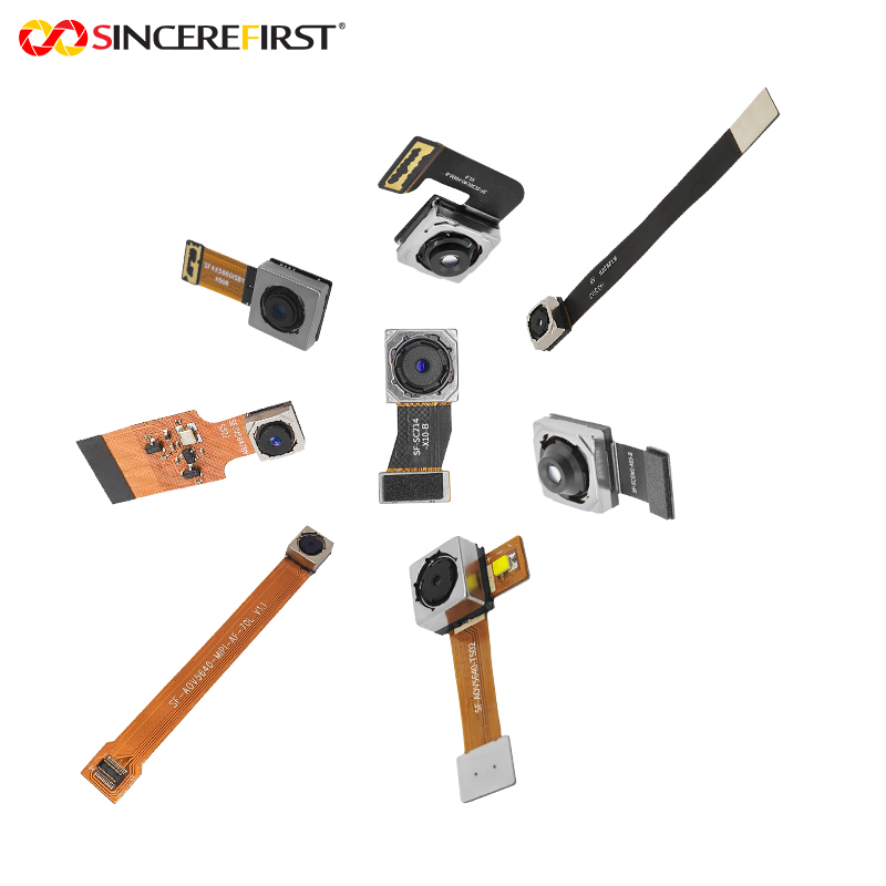
Standard modules are useful starting points, but medical products rarely stay standard through final design. Customization may include a different FPC length, connector orientation, board shape, lens barrel, viewing angle, LED arrangement, frame rate, firmware setting, or mechanical mounting method. The objective is not customization for its own sake. It is to remove compromises that would otherwise affect device size, assembly yield, imaging quality, or user experience.
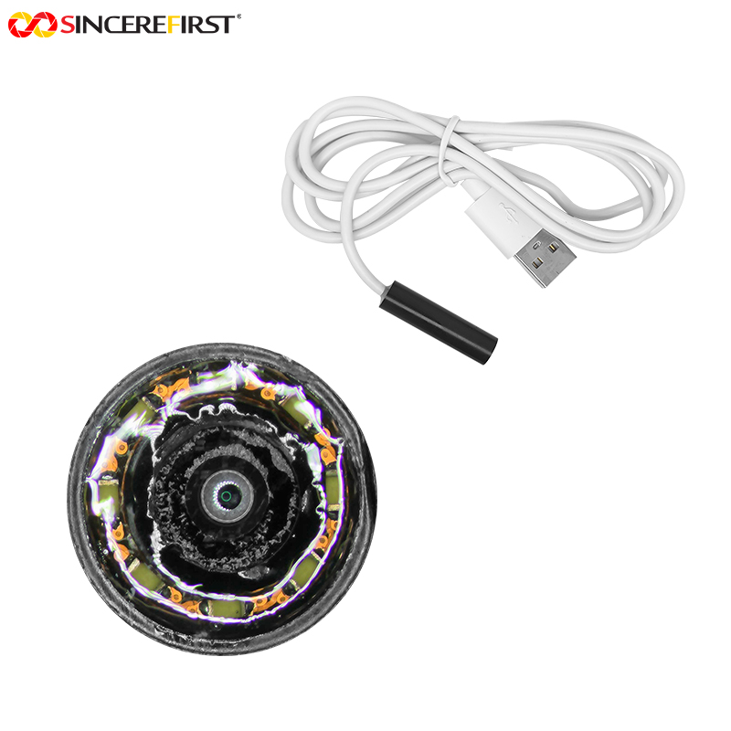
For example, an endoscope module may require a small outer diameter, side-view or forward-view configuration, controlled LED placement, and a cable designed around a specific insertion path. A dental imaging module may prioritize close-range focus, controlled illumination, and natural color. A portable diagnostic system may need low power consumption and a MIPI interface that works with an existing embedded platform.
SincereFirst supports this type of OEM development by combining camera module engineering, optical component capability, rapid sample work, and scalable manufacturing experience. The productive supplier relationship is one in which the manufacturer reviews the full integration requirement early, rather than simply shipping a catalog camera and leaving the OEM to solve optical, mechanical, and production issues alone.
How to qualify a medical imaging module supplier
A supplier evaluation should move beyond datasheets. Request representative samples and test them in the actual device architecture. Review drawings, interface timing, image tuning options, material declarations, inspection criteria, and planned production test coverage. If a custom design is required, confirm who owns the mechanical and optical review, how design changes are documented, and how pilot results transfer into volume production.
Speed is valuable, but only when sample turnaround is paired with disciplined engineering communication. Fast prototypes help teams resolve fit, focus, image quality, and firmware questions before tooling decisions become expensive. The supplier should also be able to explain realistic tolerances and trade-offs rather than promising every target at once.
The strongest medical imaging module is the one that gives the finished device dependable clinical visibility while remaining practical to integrate, validate, source, and manufacture. Define the image task first, test under real use conditions, and select a partner prepared to engineer for the full life of the product.