
A medical imaging device can fail long before clinical evaluation if its camera module was selected as a commodity part. In medical imaging, image quality is only one requirement. The module must also fit the device envelope, work predictably with illumination, transfer data reliably, tolerate the intended cleaning or sterilization process, and remain manufacturable after prototypes become production units.
For OEMs, product managers, and engineering teams, the better question is not simply, “Which camera has the highest resolution?” It is, “Which imaging architecture will deliver usable images consistently within our clinical, mechanical, electrical, and supply-chain constraints?” That distinction determines whether a project moves efficiently from proof of concept to a stable commercial device.
Medical Imaging Starts With the Use Case
The camera specification should follow the clinical task. A module for a compact otoscope, dental camera, or single-use endoscope has different priorities than one used in a laboratory analyzer, dermatology device, surgical visualization platform, or patient-monitoring product.
For close-range inspection, depth of field, controlled illumination, color fidelity, and small mechanical dimensions may matter more than extreme resolution. A larger sensor can capture more detail, but it may require a larger lens assembly, consume more power, generate more heat, and increase the processing burden. In a narrow endoscope shaft, the available diameter may set the entire optical architecture.
For external imaging systems, the requirements can shift toward low-light performance, frame rate, dynamic range, and stable color reproduction. A fast-moving procedure may benefit from higher frame rates to reduce motion blur. A system expected to reveal subtle tissue or surface differences may need carefully managed illumination and image processing, not only a higher megapixel count.
Device teams should define the working distance, field of view, target feature size, illumination geometry, expected motion, and display or recording requirements before choosing a sensor. These inputs make it possible to assess the trade-offs that actually affect image usability.
The Core Components of a Medical Imaging Module
A camera module is an integrated imaging subsystem. Its performance results from the interaction of the sensor, lens, illumination, interface, mechanical structure, and firmware. Optimizing one component in isolation can create a problem elsewhere.
Sensor Selection Is More Than Resolution
CMOS sensors are widely used in compact medical devices because they can offer high resolution, low power consumption, fast readout, and compact packaging. Yet the sensor’s pixel size, sensitivity, shutter type, signal-to-noise ratio, dynamic range, and supported frame rate may be more relevant than its headline resolution.
A rolling shutter sensor can be a practical, cost-effective choice for stationary or controlled scenes. Where the device or target moves quickly, a global shutter may better prevent geometric distortion. Low-light applications may call for larger pixels or a sensor optimized for sensitivity, while high-magnification systems may require enough resolution to preserve detail after cropping or digital zoom.
The selected sensor should also have a realistic lifecycle. For medical device manufacturers, redesign caused by sensor discontinuation is expensive. A qualified supplier should help evaluate availability, alternate sensor paths, and the implications of a component change on optics, firmware, image tuning, and validation.
Optics Define What the Sensor Can See
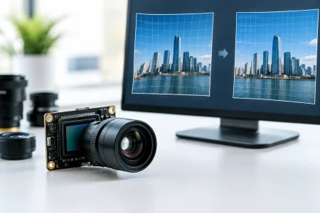
Lens selection controls field of view, focus range, distortion, sharpness, and relative illumination across the image. In close-focus devices, standard consumer-camera optics often do not provide the required performance. A lens must be designed or selected around the actual working distance and target area.
Depth of field creates a common trade-off. A smaller aperture can keep more of the target in focus, but it also reduces the light reaching the sensor. That may require stronger LEDs, longer exposure, or higher sensor gain. Each option can affect heat, power consumption, motion blur, or visible noise.
For medical applications, lens alignment is equally important. Small deviations in lens position, tilt, or focus can cause edge softness and inconsistent image performance between units. Controlled assembly, active alignment where needed, and image-based quality checks are essential when the application has tight optical tolerances.
Illumination Must Be Designed With the Camera
Many image problems attributed to a sensor are actually illumination problems. Uneven LEDs can create hotspots, shadows, glare, or insufficient brightness at the target. Reflective tissue, wet surfaces, curved cavities, and translucent materials are particularly demanding.
The illumination design should consider LED position, beam angle, diffuser materials, color temperature, thermal behavior, and drive control. High color rendering may be necessary when users need to distinguish subtle visual characteristics. In other systems, narrow-band or near-infrared illumination may support a specific inspection objective. The right choice depends on the intended use and must be evaluated at the system level.
Heat management deserves early attention. LEDs, image sensors, and processors all add thermal load, especially in compact handheld devices. Excess heat can affect user comfort, component lifetime, image noise, and long-duration operating stability.
Interfaces and Electronics Affect Product Architecture
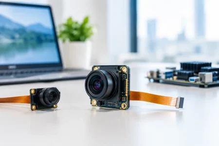
The camera interface must match the host processor, cable length, bandwidth requirements, and system power budget. MIPI CSI-2 is common in compact embedded platforms because it supports high-speed transmission with low overhead. USB camera modules can simplify development for PC-based equipment, laboratory instruments, and systems that benefit from UVC compatibility. DVP interfaces remain useful in certain lower-complexity embedded designs.
The best interface is not automatically the fastest one. MIPI integration requires compatible hardware and software resources on the host side. USB can be easier to deploy but may introduce mechanical, bandwidth, cable, or power considerations. Long cable runs, especially in larger equipment, may require additional signal-integrity planning.
Firmware and image signal processing should be discussed at the same time. Exposure control, white balance, defect-pixel correction, noise reduction, lens shading correction, and image orientation all influence the final output. A module that appears good in a supplier demonstration may require different tuning once it is installed behind a window, paired with final LEDs, or used at the intended working distance.
Mechanical Design and Reliability Are Not Secondary Details
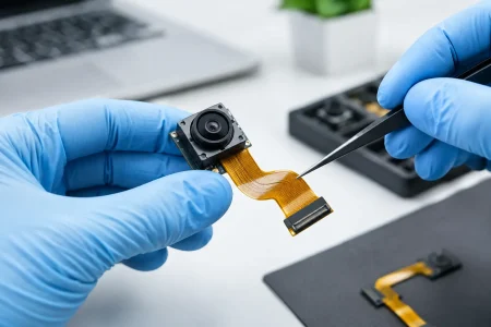
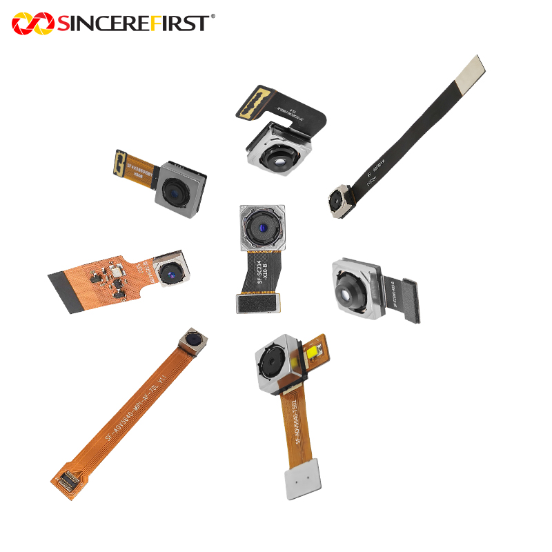
Medical device environments introduce constraints that consumer electronics rarely face. The module may need to fit into a narrow distal tip, a sealed handheld enclosure, a reusable instrument, or a disposable accessory. Connector orientation, flex cable bend radius, strain relief, shielding, mounting points, and tolerance stack-up must be designed into the assembly.
Cleaning and disinfection requirements can affect lens windows, adhesives, housings, seals, and cable materials. Reusable products may need to withstand repeated chemical exposure or defined reprocessing cycles. Single-use devices have different cost and packaging pressures. Neither approach is universally better, but each calls for deliberate material and reliability decisions.
Medical imaging modules also need consistent manufacturing control. Focus accuracy, image centering, dust contamination, cosmetic defects, electrical performance, and final functional testing should be managed with documented inspection standards. Cleanroom assembly can reduce contamination risk in sensitive optical assemblies, while traceability supports root-cause investigation if a field issue occurs.
Build for Validation and Scaled Production
A prototype that produces an attractive image is not yet a production-ready module. The transition to volume manufacturing requires stable component sourcing, defined optical alignment methods, production test coverage, packaging controls, and change-management procedures.
Early design reviews should identify parts with single-source risk, unusually tight assembly tolerances, fragile cable constructions, or test methods that cannot be repeated efficiently at scale. A qualified module manufacturer can support this work by providing sample builds, engineering feedback, optical adjustment, interface support, and a path from small pilot quantities to larger production volumes.
For products intended for regulated healthcare markets, the camera module supplier does not replace the device manufacturer’s quality and regulatory responsibilities. However, supplier documentation, process consistency, material control, and reliable technical communication can make verification and validation work far more manageable. Teams should define required records, inspection criteria, revision control, and acceptance testing before purchase orders become recurring production demand.
A Practical Specification Framework
Before requesting samples, device teams should prepare a concise imaging requirement that covers the intended use, target working distance, field of view, required resolution at the object plane, frame rate, lighting conditions, module dimensions, interface, cable length, power limits, operating environment, and anticipated annual volume. It should also identify whether the design is reusable or disposable and whether the module must fit a specific enclosure or distal-end diameter.
This level of definition prevents an all-too-common failure mode: comparing modules by megapixels and unit price while overlooking integration cost. A lower-cost standard module may be the right answer when its optics, interface, and mechanical format already fit. When the design has unusual size, illumination, cable, or performance constraints, customization can reduce downstream engineering risk and shorten the route to a manufacturable device.
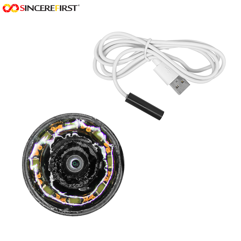
SincereFirst supports this decision process with standard and customized camera modules, including compact medical and endoscope imaging configurations. The most effective development programs begin with real application conditions, then convert those conditions into measurable optical, electrical, and production requirements.
The camera module should be treated as a controlled subsystem, not an interchangeable afterthought. Specify the image the user needs, the space the engineer has, and the production consistency the business requires. That is where a reliable medical device begins to take shape.